
Figure 4. CHAG blocks the activation of downstream signaling molecules of EGFR and inhibits EGF- induced expression of MMPs and PCNA.
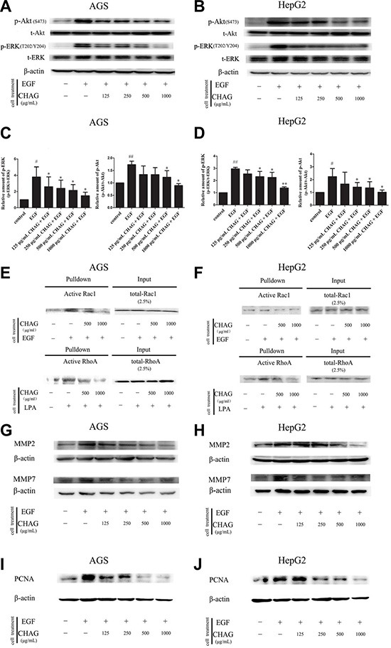
(A–D) The inhibition of CHAG on the phosphorylation/activation of Akt and ERK in AGS cells and HepG2 cells. The cells were treated same as in Figure 3 (Panel C). The cellular lysates were subjected to Western blotting with antibodies against phosphorylated Akt (p-Akt) or phosphorylated ERK (p-ERK). Total Akt (t-Akt), total ERK (t-ERK) and β-actin were detected as loading control. A and B were the representative Western blotting results of three independent experiments. C and D were results of densitometry analysis of the corresponding Western blotting results. (#P < 0.05, ##P < 0.01, compared with control group; *P < 0.05, **P < 0.01 compared with EGF group). (E and F) CHAG blocked the activation of Rac1 and RhoA in AGS and HepG2 cells. For detection of Rac1 activation, the cells were serum starved overnight, treated with EGF (100 ng/ml, 5 min), or with CHAG solutions (at concentrations of 500, 1000 μg/ml respectively) for 1 h and then with EGF (100 ng/ml, 5 min); For detection of RhoA activation, the cells were serum starved overnight, treated with LPA (1 μM, 5 min), or with CHAG solutions (at concentrations of 500, 1000 μg/ml respectively) for 1 h and then with LPA (1 μM, 5 min). The level of active Rac1 or RhoA was analyzed by ‘‘Pull-down” method. The results were representatives of three independent experiments. (G and H) Detection of the expression of MMP2 and MMP7 in AGS and HepG2 cells by Western blotting. In EGF group, the cells were treated with EGF (100 ng/ml, 24 h). In the CHAG + EGF groups, the cells were treated with CHAG at various concentrations (125, 250, 500, 1000 μg/ml respectively) and EGF (100 ng/ml) for 24 h. The cells were harvested and the lysates were subjected to Western blotting with anti-MMP2 and anti-MMP7 antibodies. (I and J) Western blotting detection of the expression of PCNA in AGS and HepG2 cells. The cells were treated same as described in panel G and H, and the lysates were probed by Western blotting with anti-PCNA antibody. The results were representatives of three independent experiments.