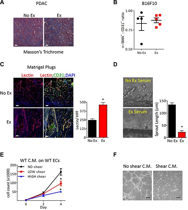
Figure 4. Exercise-induced tumor vascular remodeling is mediated in an endothelial cell autonomous manner.

(A) Representative image of Masson's trichome staining of PDAC-4662 tumors from exercised or non-exercised mice. (B) Quantification of for α-SMA and CD31 immunofluorescence of B16F10 tumors from exercised or non-exercised mice. Graph shows the average α-SMA:CD31 ratio +/− S.E.M for individual tumors, n = 4–5, bar = 100 μm. (C) Representative images and quantification of isolectin-B4 and CD31 in Matrigel plugs. Matrigel plugs with ECs harvested from mice after 7 days of exercise or non-exercise control were immunostained for isolectin-B4 (red), CD31 (green), and DAPI (blue), n = 4, * p < 0.05, Bars = 50 μm. (D) Representative images of EC sprouting in a microfluidic device in vitro after exposure to circulating media containing serum from exercised or non-exercised mice. Average sprout length was quantified after 24 hours, shown as the mean +/− S.E.M., *p < 0.05, n = 3. (E) Proliferation of naïve ECs at the indicated days in media conditioned by ECs after exposure to either no, low or high shear stress for 24 hours. Data are shown as mean +/− S.E.M., *p < 0.05, n = 3. (F) Images of Matrigel tube formation by naïve ECs in conditioned media harvested from ECs exposed to either no or high shear stress, Bar = 50 μm.
