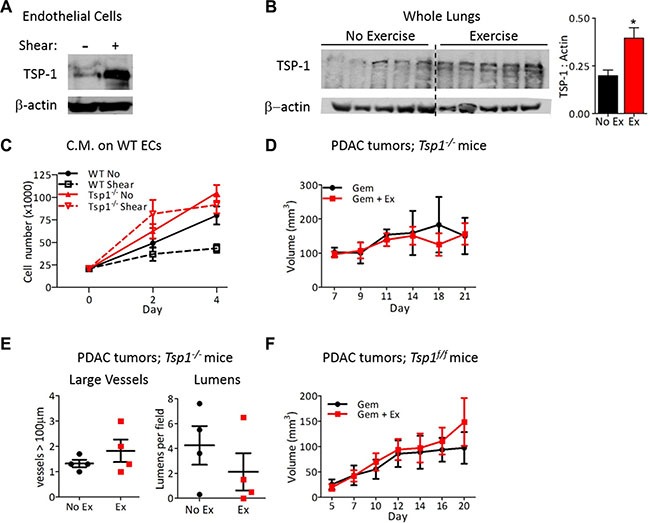
Figure 6. Endothelial-derived TSP-1 is critical for increased chemotherapeutic efficacy by shear stress.

(A) Immunoblots for TSP-1 expression by mouse lung ECs after exposure to 0 or 24 hours of shear stress (8 dynes/cm2). Actin was probed as a loading control. (B) Immunoblot for TSP-1 expression by lungs from wild-type mice exercised for two weeks with actin as a loading control. Each lane is a lung from one mouse. TSP-1 expression is quantified relative to actin, n = 5, *p < 0.05. (C) Proliferation of wild type or Tsp1−/− ECs in conditioned media from wild type or Tsp1−/− mLung ECs after exposure to 0 or 24 hours of shear stress. Data are represented as mean +/− S.E.M., n = 3, *p < 0.05. (D) Tumor growth in Tsp1−/− mice. PDAC-4662 cells were inoculated in the flanks of Tsp1−/− mice. Mice were treated with gemcitabine with or without exercise by the same protocol used in wild type mice for Figure 2. Tumor volumes shown as mean +/− S.E.M., n = 4, not significant. (E) Quantification of vascular normalization on tumors from Tsp1−/− mice. PDAC-4662 tumors from Tsp1−/− mice treated with exercise or non-exercise control were assessed by immunofluorescence staining for vessels >100 μm and the number of visible lumens per field. 5 sections per tumor were quantified and used to obtain one value per tumor. Each marker represents one tumor, n = 4, not significant. (F) PDAC-4662 flank tumor growth in VE-cadherin-Cre; Tsp1fl/fl mice. PDAC-4662 cells were inoculated into VE-cadherin-Cre; Tsp1fl/fl mice. When tumors were palpable, mice were treated with gemcitabine with or without exercise. Data are average tumor volumes +/−S.E.M. on the indicated days, n = 5, not significant.
