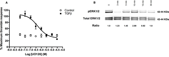
Figure 4. TGFβ-ERK1/2 signalling is also involved in regulation of PAR-1 expression in A549 cells.

Panel A. Dose-inhibition curve in A549 cells exposed to TGFβ (1 ng/ml) with varying concentrations of MEK1/2 inhibitor, UO126, for 24 hours and then stimulated with thrombin (30 nM). Each data point represents the mean +/− SEM of 3-4 replicate wells. Panel B. Representative immunoblot of ERK1/2 phosphorylation in A549 cells incubated with TGFβ (1 ng/ml) for up to 30 minutes with corresponding pERK/total ERK ratios derived from densitometry analysis.
