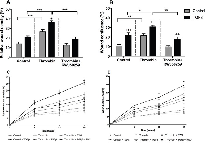
Figure 7. The effect of TGFβ treatment on A549 lung adenocarcinoma cells migration.

A549 cells were incubated with or without TGFβ (1 ng/ml) for 24 hours and pre-treated with PAR-1 inhibitor, RWJ58259 (3 μM) for 30 minutes. Homogenous scratch wounds were introduced using WoundMaker (Essen Bioscience, UK) and cells immediately stimulated with thrombin (10 nM). Wound closure was monitored for up to 24 hours and data analysed using Incucyte Zoom Live Cell Imaging System (Essen Bioscience, UK). Panel A. Difference in wound cell density and Panel B. Wound confluence at 24 hours. Panel C and D. Time-course of wound closure with data collected at 6, 12 and 18 hours with control and control + TGFβ denoted by dashed lines, thrombin and thrombin + TGFβ denoted by continuous lines and thrombin + RWJ58259 and thrombin + TGFβ + RWJ58259 denoted by dotted lines. Data are plotted as mean +/− SEM of n=5 replicate wells from 2 independent experiments; Two-way ANOVA, +p<0.05, ++p<0.01, +++p<0.001 in comparison to control, *p<0.05, **p<0.01, ***p<0.001 comparison between stimulations.
