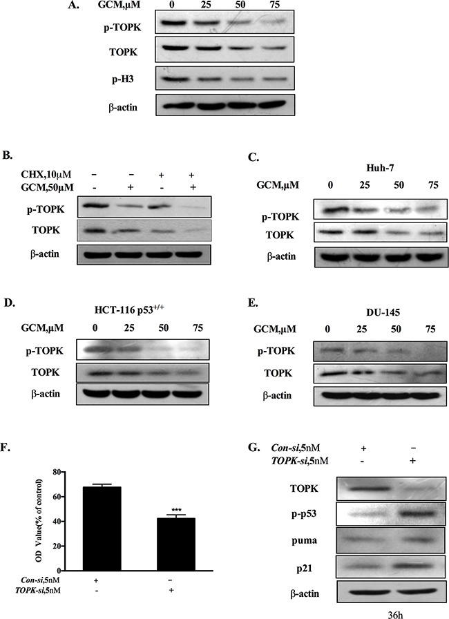
Figure 3. Activation of p53 by GCM is attributed to suppression of TOPK.

A. GCM inhibited TOPK in HepG2 cells. The cells were treated with various concentrations of GCM for 24 h and the changes of phospho-, total TOPK and its substrate histone H3 phosphorylation were determined by western blotting. B. GCM promoted TOPK degradation in HepG2 cells. The cells were treated with 50μM of GCM in the presence or absence of CHX and the expression of TOPK was analyzed by western blotting. C. GCM inhibited TOPK in Huh-7 cells. D. GCM inhibited TOPK in HCT-116 cells. E. GCM inhibited TOPK in DU145 cells. F. Overall inhibitory effects of TOPK knockdown on HepG2 cells measured by crystal violet staining. G. TOPK knockdown activated p53 in HepG2 cells. The cells were transfected with 5 nmol/L of TOPK siRNA or non-targeting siRNA for 36 h and the changes of p-p53, p21 and puma were analyzed by western blotting.
