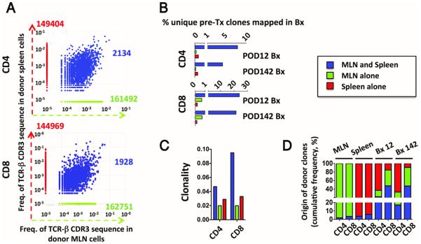
Figure 6. Origin of early detected GvH clones in Patient 13.
(A) Scatter plots showing donor clones identified in the mesenteric lymph nodes (MLN) alone (green), in the spleen alone (red) and those found in both the mesenteric lymph nodes and spleen (blue) in Patient 13. (B) The bar graphs depict the proportion of the three donor-derived cell subsets (green, red and blue) also detected in the post-transplant intestinal biopsies with a sizeable donor population. (C) Clonality of CD4 and CD8 clones from the three subsets. (D) Proportion of blue, green and red clones among the total donor clones in the MLN, spleen and post-transplant biopsies.

