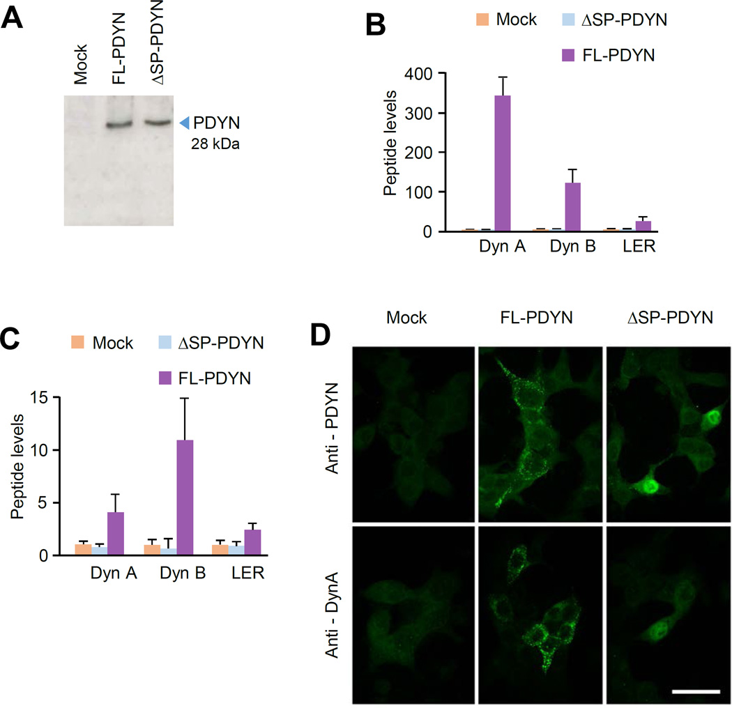
FIGURE 3. Processing and subcellular localization of ectopically expressed ΔSP- and FL-PDYN in RINm-5F cells.
A. ΔSP- and FL-PDYN proteins analyzed by western blotting with anti-PDYN CTF antibody in RINm-5F transfected cells.
B, C. The levels of prodynorphin derived dynorphin A (Dyn A), dynorphin B (Dyn B) and Leu-enkephalin-Arg (LER) in the RINm-5F transfected cells (B) and in the culture media (C) analyzed by RIA. The cells were transfected with pCMV vector (mock), pCMV-ΔSP-PDYN or pCMV-FL-PDYN. Data were normalized to the levels in mock transfected cells and are shown as mean ± SEM; n = 3 experiments.
D. Subcellular localization of ΔSP-PDYN protein in RINm-5F cells observed under confocal fluorescence microscopy. Transfected cultures were immunolabeled with antibodies against PDYN CTF or dynorphin A. Images were taken with × 100 objective and × 5 electron zoom factor. Scale bar, 30 µm.

