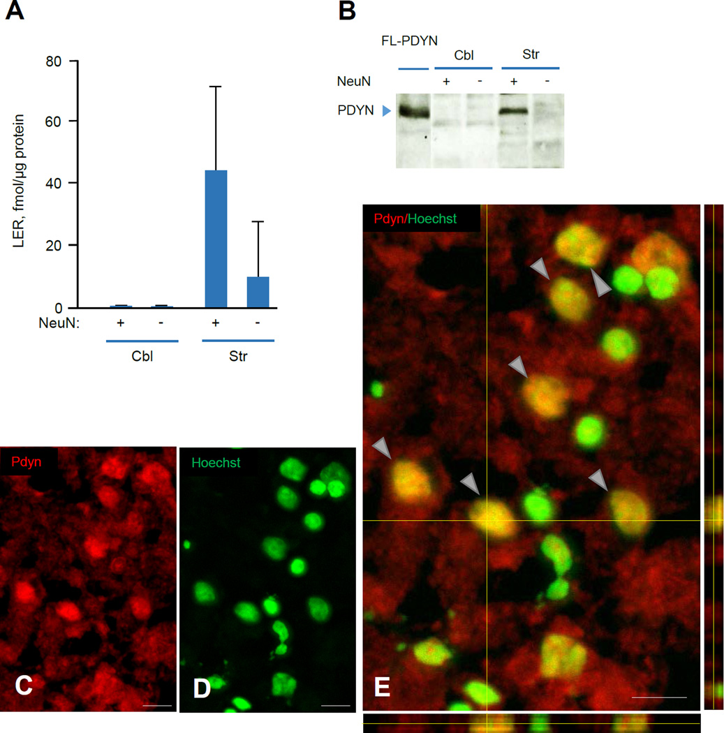
FIGURE 4. ΔSP-PDYN is targeted to the neuronal nuclei in human striatum.
A. The levels of PDYN derived peptide LER in neuronal and non-neuronal nuclei isolated by FANS from human striatal (Str) and cerebellar (Cbl) tissue samples. LER, a PDYN marker was analyzed by RIA. The amount of the liberated LER in both neuronal and non-neuronal nuclei prepared from cerebellum was below the detection limit. Equal amount of neuronal and non-neuronal nuclei (106 nuclei) isolated from each tissue sample were analyzed. Data is shown as mean ± SEM, n = 2 subjects. Data was normalized to 1 µg of protein.
B. PDYN proteins analyzed by western blotting in neuronal and non-neuronal nuclei isolated by FANS from human striatal (Str) and cerebellar (Cbl) tissue samples. Equal amount of neuronal and non-neuronal nuclei (106 nuclei) isolated from each tissue sample were analyzed. Ectopically expressed in RINm-5F cells FL-PDYN was used as a positive control, producing a single band with molecular size of 28 kDa that corresponded to unprocessed PDYN protein.
C–E. PDYN immunoreactivity was detected in the neuronal cell nuclei and cytoplasm in the human caudate nucleus. The brain sections were stained for ΔSP-PDYN protein with anti-PDYN CTF antibody (red) (C) and for nuclei with Hoechst (green) (D). Representative 3D confocal reconstruction projections demonstrates double labeling (yellow) of neuronal nuclei (arrows) (E) indicating nuclear localization of PDYN isoform. Confocal images of Z-stacks of 24 images acquired with a depth interval of 0.5 µm of the human caudate nucleus. Analysis shows that PDYN is present in the nucleus. The center image is the X-Y view, the images below and right are orthogonal projections in the X-Z and Y-Z planes (cross section at the yellow line). n = 4 subjects. Images were taken with 20 × /0.95W XLUMPlanFI water objective. Scale bar, 20 µm.

