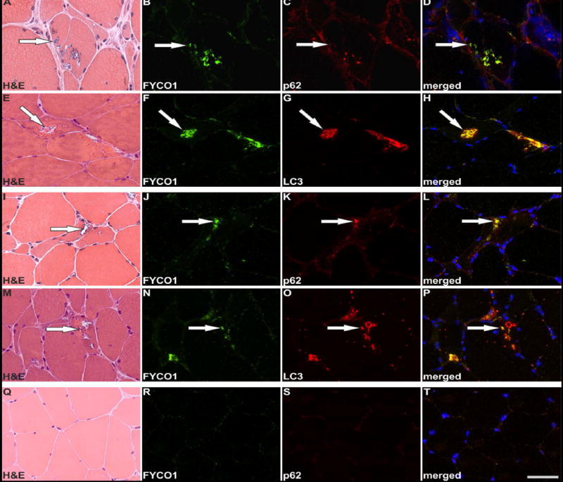
Figure 3. Co-localization of FYCO1, p62 and LC3 in RVs of sIBM patients.

Serial skeletal muscle sections from two sIBM patients (patient 1: A-H, patient 2: I-P) and from a healthy control (Q-T) were stained with H&E and double-immunostained with primary antibodies directed against FYCO1 (green) and p62 or LC3 (red). Nuclei are stained with DAPI (blue). For each sIBM patient two different RV containing areas of the muscle samples are displayed. All RVs show a strong immunoreactivity for FYCO1, p62 and LC3. The co-localization of FYCO1 with p62 LC3 is indicated by yellow in the merged images. Scale bar = 50 μm.
