
Figure 4. Validation of proteomic findings by immunolocalization studies I.
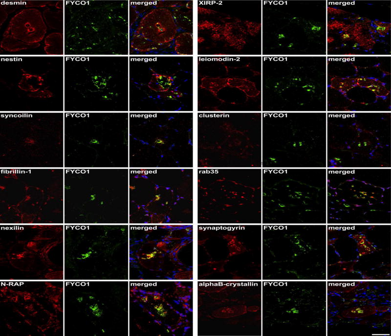
Serial sections from two sIBM patients were stained with H&E and double-immunostained with antibodies recognizing desmin, nestin, syncoilin, fibrillin-1, nexilin, N-RAP, XIRP-2, leiomodin-2, clusterin, rab35, synaptogyrin, and alphaB-crystallin. All proteins showed an accumulation in RV samples (red) and FYCO1 (green) as a positive control to localize RVs. Nuclei are stained with DAPI (blue). Increased immunoreactivity was observed with all proteins as indicated by yellow in the merged image. Scale bar = 50 μm.