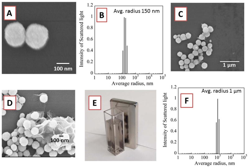
Fig. 2.
Morphology of Fe3O4 nanoparticles: (A) SEM image showing two Fe3O4 nanoparticles and (B) DLS of Fe3O4 nanoparticles with average diameter 300 nm. (C & D) SEM Images of Fe3O4 on surface of GO sheets showing morphology of Fe3O4@GO, (E) Magnetic attraction of Fe3O4@GO nanoparticles in the cuvette to the magnet on the right (F) DLS of Fe3O4@GO composite.

