Fig. 2.

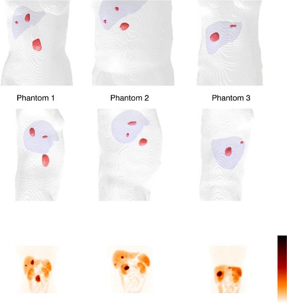
Tumour shapes and positions in the phantoms. The parts of the body outlines and the liver are included as anatomical references. The bottom row shows total intensity projections of the corresponding simulated SPECT images at 24 h p.i

Tumour shapes and positions in the phantoms. The parts of the body outlines and the liver are included as anatomical references. The bottom row shows total intensity projections of the corresponding simulated SPECT images at 24 h p.i