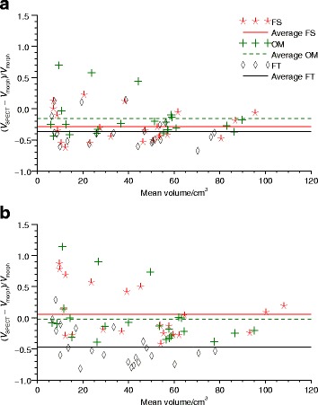
Fig. 7.

Volume differences between manual delineation in morphological images and SPECT image segmentation. The three methods FT, OM, and FS are used. SPECT images were reconstructed with a ASR and b AS. Differences are expressed as the volume differences divided by the volume derived from morphological images and are shown as function of the average volumes derived from SPECT and morphological images
