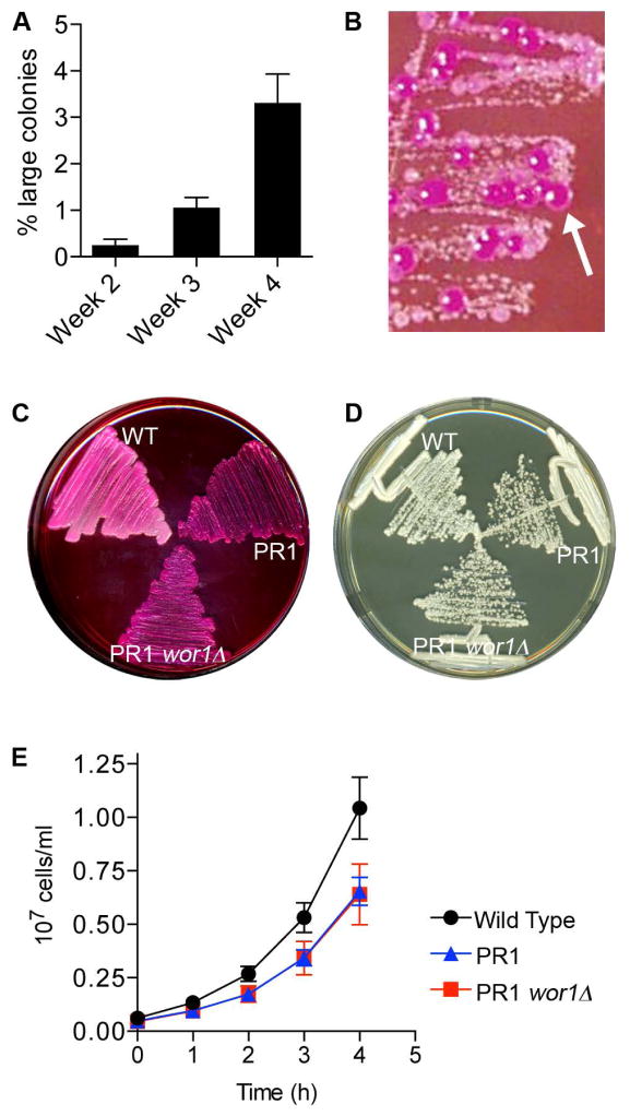
Figure 7. Pseudorevertants show increased Phloxine B staining, but not other characteristics of Opaque cells.
A. The cyr1Δ strain CR216 was incubated at room temperature on YPD agar medium for the indicated time, restreaked onto fresh medium, and then the resulting colonies were assessed to determine the percent of faster-growing large colonies rather than the expected small colonies. The results represent the average for 6 colonies aged for two weeks, 59 colonies aged for three weeks, and 91 colonies aged for four weeks.
B. The cyr1Δ cells (CR216) were incubated 3 weeks on YPD agar plates and then streaked onto a YPD plate containing 45 μg ml−1 Phloxine B. Larger pseudorevertant colonies were observed to stain magenta (arrow), similar to that expected for cells in the Opaque phase.
C. Wild type (DIC185), a cyr1Δ pseudorevertant (PR1) derived from strain CR216, and its wor1Δ derivative (PR1-wor1Δ) were streaked onto YPD agar plate containing 45 μg ml−1 Phloxine B, showing that deletion of WOR1 did not change Phloxine B staining of the pseudorevertant.
D. The strains described in panel C were streaked for single colonies on YPD and incubated at 30° C for 2 days.
E. The strains described in panel C were grown in liquid YPD media at 30° C. Panels D and E show that deletion of WOR1 does not affect the growth rate of the pseudorevertant strain PR1.

