
FIGURE 6.
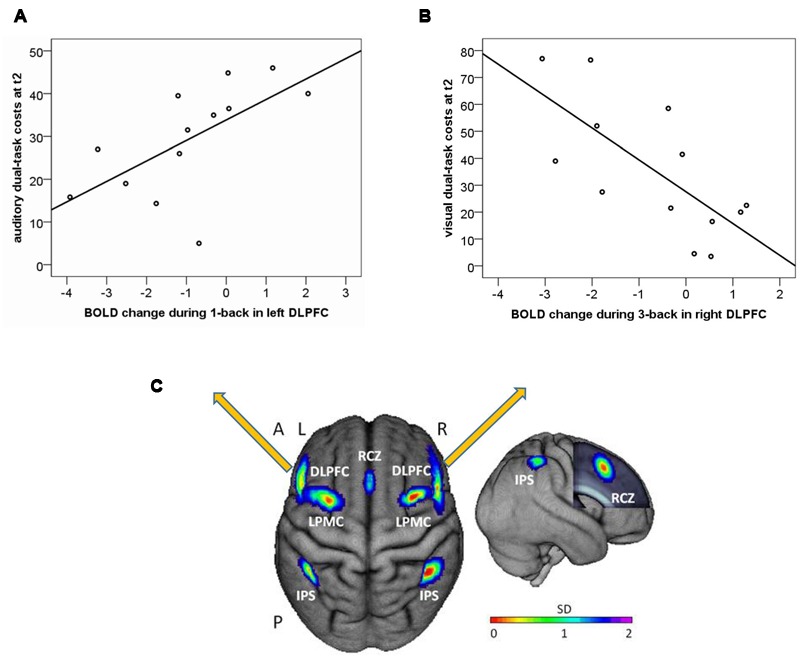
Correlation of one-back BOLD change from T1 to T2 (arbitrary units) and auditory dual-task costs at post-test (T2) for (A) left dorsolateral prefrontal cortex (DLPFC); and (B) right dorsolateral prefrontal cortex (DLPFC); (C) Location of literature-based probabilistic ROIs of the WM network. Left item: dorsal view of the ROIs overlaid onto the surface of the sample mean brain. Right item: right lateral view. The frontal lobe was cut to display the mid-sagittal ROI. RCZ, rostral cingulate zone; DLPFC, dorsolateral prefrontal cortex; LPMC, lateral premotor cortex; IPS, intraparietal sulcus; A, anterior; P, posterior; L, left; R, right; SD, standard deviation.