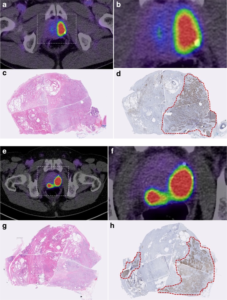
Fig. 6.
Comparison of virtual whole mount histopathology (H&E and PSMA-immunostaining) and PSMA PET-findings. Transaxial PET/CT-scan of patient 1 (a, b, e, f) and corresponding histopathology of the subsequent prostatectomy specimen; H&E staining (c, g); PSMA-immunostaining with outlined tumor contours in red (d, h)

