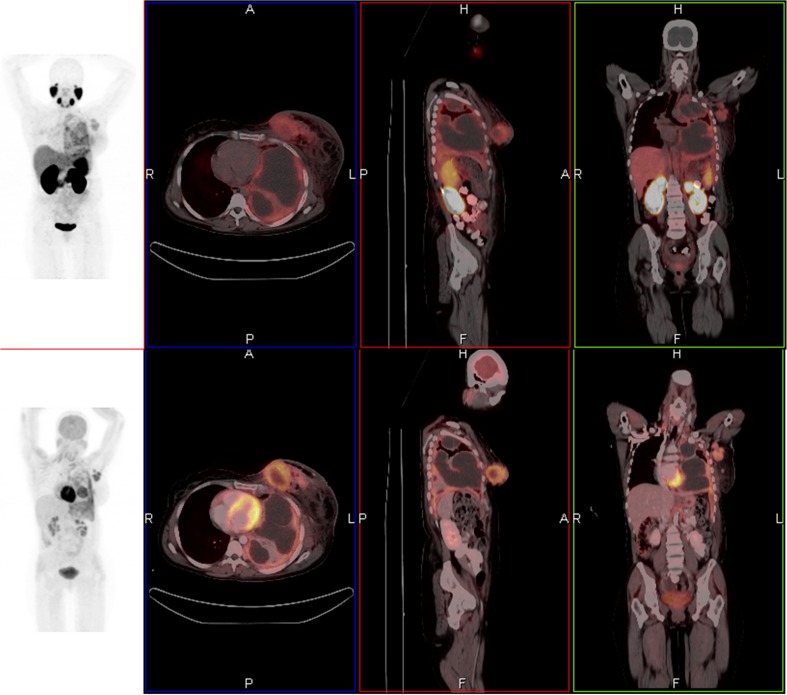
Fig. 1.
A 42-year-old female with metastatic breast carcinoma who underwent 68Ga-PSMA and 18F-FDG PET/CT. Axial, coronal, and sagittal fused 68Ga-PSMA PET/CT images demonstrated primary left breast cancer, axillary nodal and left pleural metastases (a). Avidity is slightly intense on 18F-FDG PET/CT images (b). Maximum-intensity-projection PET gives overview of all lesions (c, d)

