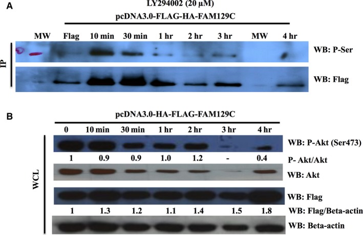
Figure 6.

Effect of PI3K inhibition on BCNP1 phosphorylation at serine residues. (A) IP and (B) WB analyses were performed with the indicated antibodies. HEK293 cells were transfected with Flag‐HA‐BCNP1. After expression, cells were treated with indicated PI3K inhibitors and then incubated for the indicated period of time. Transiently expressing Flag‐HA‐BCNP1 HEK293 cells were treated with PI3K inhibitor LY294002 (20 μM) for 10 min., 30 min., 1, 2, 3, and 4 hrs. HA, haemagglutinin A; WCL, whole cell lysate; IP, immunoprecipitation; WB, western blot; MW, molecular weight marker.
