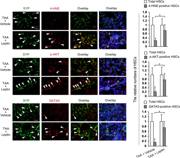
Figure 5.

Leptin increases the levels of 4‐HNE, p‐AKT, and GATA3 in HSCs in ob/ob mouse model of TAA‐induced liver injury. Two groups of ob/ob mice (6 mice/each group) were received TAA (200 μg/g body weight, two times a week) plus vehicle (TAA+Vehicle) or TAA plus leptin (1 μg/g body weight, once per day) (TAA+Leptin) by intraperitoneal injection (i.p.) for 4‐week. Double fluorescence staining on the section of liver was performed for detecting 4‐HNE‐, p‐AKT‐, or GATA3‐positive HSCs by using the respective primary antibody plus primary antibody against synaptophysin (SYP, a marker for quiescent and activated HSCs) and subsequently the DyLight594‐conjugated secondary antibody and DyLight488‐conjugated secondary antibody. The nuclei were counterstained with Hoechst 33342 (blue fluorescence). The representative images were captured with the fluorescence microscope, scale bar 25 μm. Arrowheads indicated examples of positively stained cells. The total HSCs (SYP‐positive HSCs, green fluorescence) and 4‐HNE‐, p‐AKT‐, or GATA3‐positive HSCs (red fluorescence) were counted in six randomly chosen fields at 100‐fold magnification and the values were expressed as fold changes relative to the respective total HSCs (empty column). The values were shown as a histogram on the right panel. *P < 0.05.
