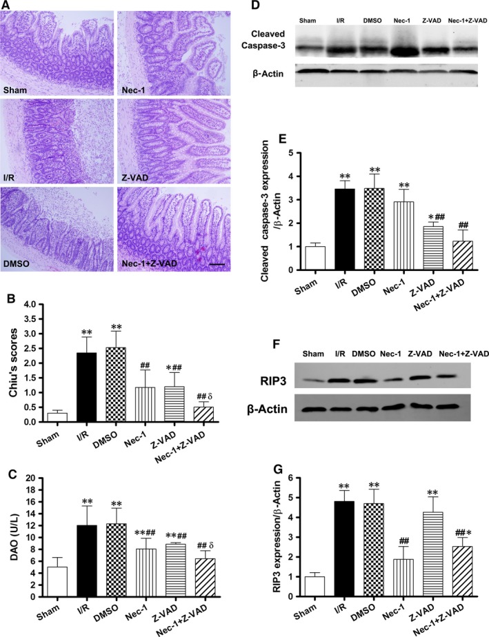
Figure 4.

Increased protection from intestinal I/R injury by the combined blockade of necroptosis and apoptosis after 1 hr of ischaemia/24 hrs of reperfusion in vivo. (A) Histopathologic changes of the intestinal mucosa. Haematoxylin and eosin stained small intestine. Magnification is ×200, bar denotes 100 mm. (B) Injury scores of the intestinal mucosa morphology. (C) Intestinal cellular injury was evaluated by serum DAO activity. (D and E) Z‐VAD with/without Nec‐1 treatment decreased the caspase‐3 cleavage. (F and G) Treatment with Z‐VAD alone had no effect on RIP3 up‐regulation. Caspase inhibition shifted intestinal I/R‐induced epithelial cell death from apoptosis to necroptosis. The images are representative for each group. The data are shown as the means ± S.D. (n = 8 per group). *P < 0.05, **P < 0.01 compared with sham group, ## P < 0.01 compared with I/R group and DMSO group, δ P < 0.05 compared with Nec‐1 group and Z‐VAD group.
