Figure 1.

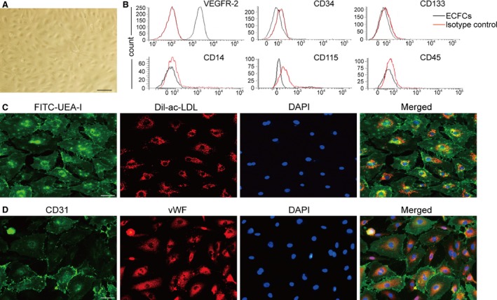
Characterization of ECFCs derived from human peripheral blood. (A) After 14–28 days of culture, cells presented a typical cobblestone shape as shown by a phase‐contrast inverted microscope. (B) FACS analysis found that most of the cells expressed VEGFR‐2, a small percentage of cells expressed CD34 and few expressed CD133, CD14, CD45 and CD115 (isotype control IgG staining shown in shaded red). (C) Testing of DiI‐ac‐LDL endocytose and FITC‐UEA‐1 binding showed that the cells were double positive. (D) Immunofluorescence staining demonstrated that the cells expressed CD31 and vWF, counterstaining with DAPI for nucleus, scale bar: 50 μm.
