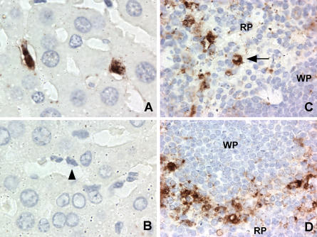
Figure 1. MNV-1-Specific Staining In Vivo Occurs in Cells of the MΦ Lineage.
Immunohistochemistry was performed on liver (A and B) and spleen (C and D) sections from STAT1−/− mice 2 d after oral infection. MNV-1-specific staining was seen in Kupffer cells of infected livers when probed with MNV-1 immune (A) but not preimmune (B) serum. A selected Kupffer cell lining the sinusoid is indicated by an arrowhead. MNV-1-specific staining consistent with MΦ was seen in red pulp (C) and marginal zone (D) in the spleen. The arrow indicates a cell with MΦ morphology. No staining was observed in tissues from mice infected for 1 d, in infected tissues incubated with preimmune serum, or in mock-infected tissues incubated with immune serum. RP, red pulp; WP, white pulp.

