Figure 1.

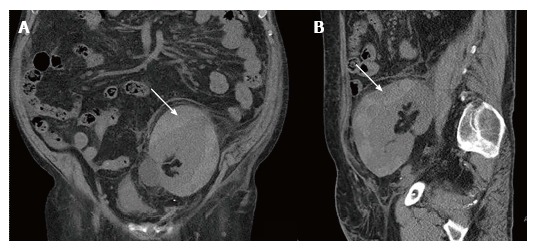
Computed tomography without intravenous contrast of the transplanted kidney. A: Coronal view; B: Sagittal view. A subscapular hematoma 12 cm × 2.5 cm in size was compressing the transplanted kidney (arrows).

Computed tomography without intravenous contrast of the transplanted kidney. A: Coronal view; B: Sagittal view. A subscapular hematoma 12 cm × 2.5 cm in size was compressing the transplanted kidney (arrows).