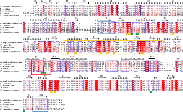
Figure 7. Sequence alignments of aldehyde dehydrogenases towards the aromatic aldehydes with broad specificity.
Identical residues are shown in white letters with a red background, similar residues are shown in red letters with a white background, and varied residues are shown in black letters. The predicted secondary structure is shown at the top of the alignment. α-Helices are represented as helices and β-strands as arrows, while β-turns are identified by ‘TT’ and 310-helices by η. The core regions of the catalytic domain, NAD+ binding domain, and oligomerization domain are boxed by red, orange, and green rectangles, respectively. Amino acid residues involved in catalysis, NAD+ binding, and SAL binding are labeled by a red star, orange square, and green square, respectively. Aldehyde dehydrogenases are from A. naphthalenivorans (F5Z5S7), P. putida (Q1XGL7), C. glutamicum (Q8NMB0), B. linens (A0A142NN86), and G. thermoleovorans (A0A098L0N3).

