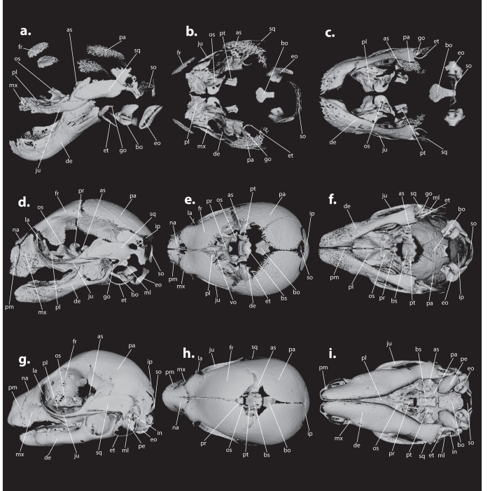
Figure 2. One plate with three different stages of cranial bone development in Petrogale penicillata.
The left lateral side of the skull is shown in a,d, and g, the dorsal view in b,e, and h, and the ventral view in c,f, and i. Images were created using MeshLab 1.3.3 and Adobe Illustrator CS6. a,b, and c represent ZMB_EMB_MA587, the earliest of the three stages; d,e, and f represent ZMB_EMB_MA590, the intermediate stage; and g,h, and i represent ZMB_EMB_MA593A, the most advanced of the three stages illustrated here. Plates of the other four species can be found in the Suppl. Figs 3, 4, 5, and 6. Abbreviations (for all figures): as (alisphenoid), bo (basioccipital), bs (basisphenoid), de (dentary), eo (exoccipital), et (ectotympanic), fr (frontal), go (goniale), in (incus), ip (interparietal), ju (jugal), la (lacrimal), ml (malleus), mx (maxilla), na (nasal), os (orbitosphenoid), pa (parietal), pe (petrosal), pl (palatine), pm (premaxilla), pr (presphenoid), pt (pterygoid), so (supraoccipital), sq (squamosal), vo (vomer).

