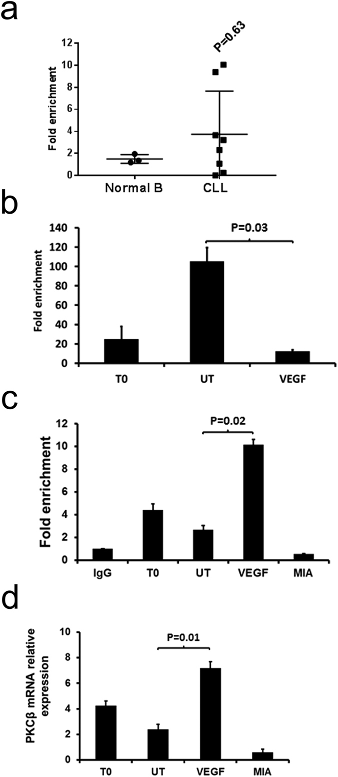
Figure 5. VEGF stimulates SP1 association with the PRKCB promoter sequence in CLL cells.

5 × 10⁶ CLL cells were used directly (T0) or cultured overnight in the absence (UT) or presence of 100 ng/mL VEGF or 200 nM mithramycin (MIA). (a) ChIP analysis of STAT3 association with the PRKCB promoter in CLL and normal B cells in individual samples. The mean ± SD of these experiments is displayed. Statistical analysis was performed using a Mann-Whitney U-test. (b) ChIP analysis of STAT3 association with the PRKCB promoter in CLL cells incubated overnight ± VEGF. (c) ChIP analysis of SP1 association with the PRKCB promoter using the same CLL samples as in part (b). IgG is the immunoprecipitation control. (d) qRT-PCR analysis of PKCβII mRNA levels in CLL cells measured in comparison to RNApolII. For ChIP analyses PRKCB promoter sequences associated with STAT3/SP1 were detected by qPCR and are presented as fold enrichment compared to the PRKCB promoter sequences associated with the non-specific IgG immunoprecipitation control. In parts (b,c and d) the data presented represent the mean ± SE of n = 3 experiments using CLL cells from different patients. Statistical analysis was performed using a students t-test for paired data.
