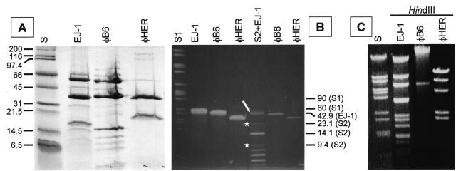
FIG. 2.
Characterization of phages φB6 and φHER and their DNAs. (A) SDS-PAGE (15% gels) showing the structural virion proteins. The proteins of EJ-1 are shown for comparison. The molecular mass (in kilodaltons) of the standards (lane S) is indicated at the left. (B) Pulsed-field gel electrophoresis of proteinase K-treated phage DNAs. Two different amounts of DNA were loaded in the same gel. In the well labeled S2+EJ-1, EJ-1 DNA (open arrow) was mixed with a size standard mixture (S2) consisting of λ DNA digested with either HindIII (white stars) or BstEII. The size (in kilobases) of several DNA bands is indicated at the right. S1, SmaI-digested S. pneumoniae R6 DNA. (C) Agarose (0.7%) gel electrophoresis of phage DNAs digested with HindIII. S, BstEII-digested λ DNA.

