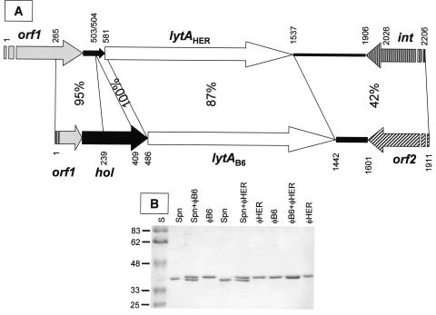
FIG. 4.
Schematic representation of the lysin genes from phages φHER and φB6 and their flanking regions, and analysis of the purified S. mitis phage lysins. (A) Arrows show the genes and their direction of transcription. The nucleotide identity between different regions of both sequences is indicated. Broken arrows correspond to incomplete open reading frames. The putative deleted holin gene from φHER is shown as a narrow, solid arrow. The nucleotide positions are also indicated. (B) The purified LytA amidases from S. pneumoniae R6, φB6, and φHER were analyzed in SDS-10% polyacrylamide gels alone or in combination. The molecular mass (in kilodaltons) of the standards (S) is indicated at the left.

