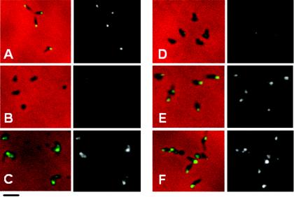
FIG. 7.
Phase-contrast-immunofluorescence analysis of P1 localization in wild-type M. pneumoniae (A), mutant M6 (B), and mutant M6 producing HMW1 Δ1-4 (C), HMW1 Δ4 (D), HMW1 Δ1 (E), or reHMW1 (F). For each pair of images, the merged phase-contrast and immunofluorescence images are shown on the left, and the corresponding immunofluorescence images alone are shown on the right. Bar, 2.0 μm.

