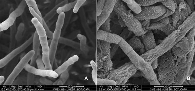
Fig. 1.

Scanning electron microscopy of Pythium insidiosum from control group (A) and treated with MFC of bark extract of S. adstringens (B). Observe the cylindrical morphology of hyphae and smooth surface of cell wall from control group, while the hyphae treated with bark extract show rough surface of cell wall, high amount of granular material and release of anamorphic material
