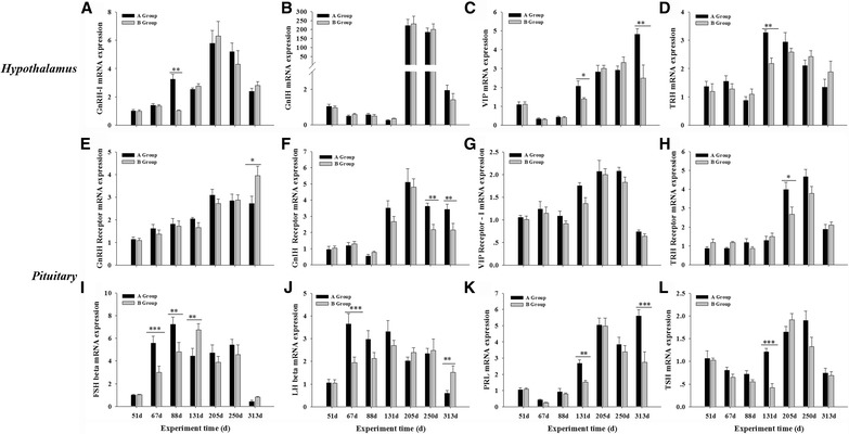
Fig. 5.

Hypothalamic (a-d) and pituitary (e-l) mRNA levels in Yangzhou goose ganders under two artificial photoperiods. Graphs a to d represent mRNA expression levels of hypothalamic releasing hormones GnRH-I, GnIH, VIP and TRH, respectively, while e to h their corresponding receptors in the pituitary gland, and i to l the FSH, LH, PRL and TSH pituitary hormones or beta subunits wherever applicable. Each value represents the average of data from eight independent culture experiments. Data are shown as mean values ± standard error of the mean. *, ** and *** indicate statistical significance based on P < 0.05, P < 0.01and P < 0.001, respectively, between the treatments
