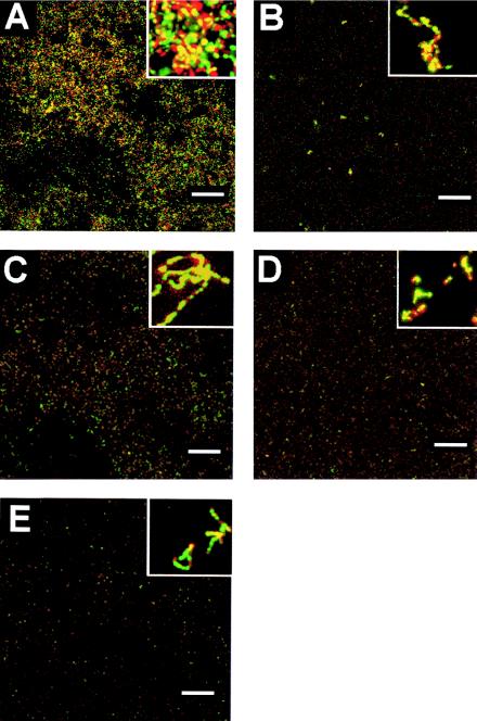
FIG. 8.
Confocal scanning laser microscopic analysis of S. gordonii biofilms in a flow-cell system. S. gordonii Challis 2 (A), nosX::Tn917-lac (B), nosX::Kanr (C), qor1::Kanr (D), and qor2::Kanr (E) strains were grown in BM in a BST FC71 flow cell unit (flow rate, 180 μl/min; initial inoculum, A660 = 0.1) over 18 h at 37°C. Live cells were stained with SYTO-9 (green), and dead cells were stained with propidium iodide (red); cells were then examined by confocal microscopy. A representative image from a set of randomly selected x-y stacks is shown for each strain. Bars, 100 μm. The small inset in each panel represents a 10× magnification of the panel.

