
Fig. 4.
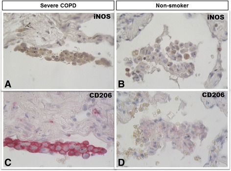
Immunohistochemistry of M1 and M2 alveolar macrophages in lung tissue. M1 (iNOS+) and M2 (CD206+) expression in clusters of alveolar macrophages in consecutive lung sections from a patient with severe COPD (panels a and c) and from a non-smoker (panels b and d). iNOS immunoreactivity appears as a brown diffuse cytoplasmic granular pattern (panel a), while CD206 immunoreactivity appears as a red linear pattern around the cellular membrane (panel c). In the smoker with severe COPD both M1 (iNOS+) (panel a) and M2 (CD206+) (panel c) immunoreactivity was present in the same cluster of alveolar macrophages. The alveolar macrophages in the non-smoking subject (panels B and D) were mostly negative for either stains. Immunostaining with anti-iNOS (in brown) and anti-CD206 (in red). Original magnification: X 400