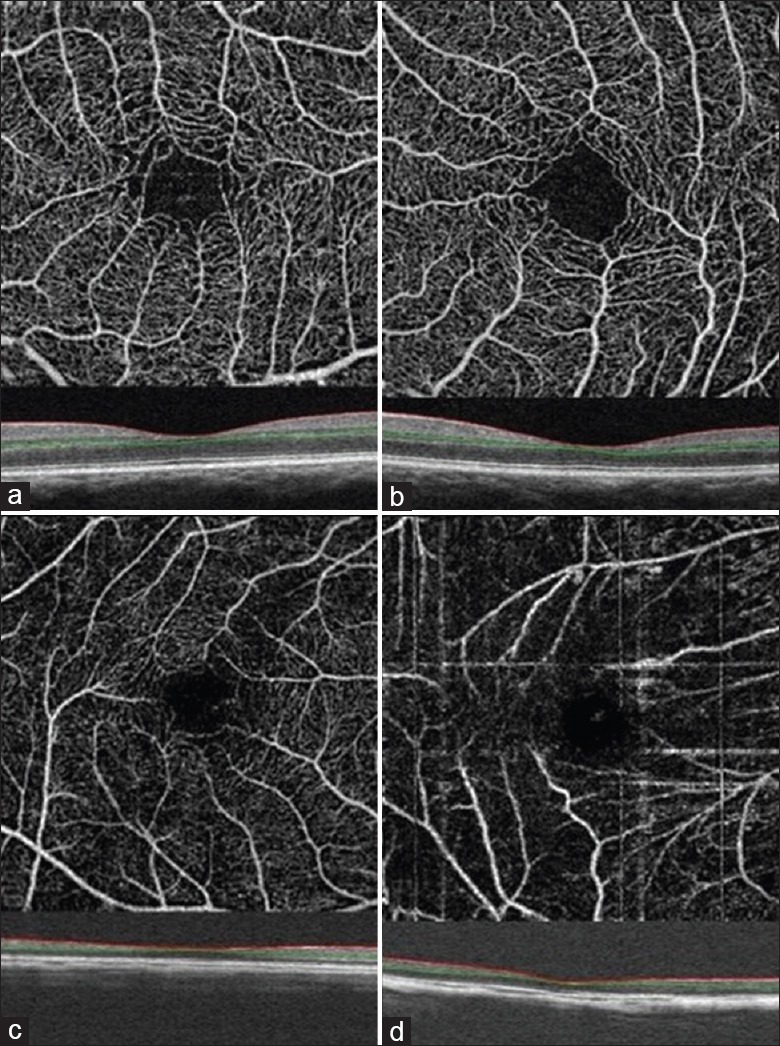
Figure 5.

Optical coherence tomography angiography images of superficial macular vessels in eyes with various axial lengths: the axial length is 23.30 mm (a) 26.11 mm (b) 29.47 mm (c) 34.62 mm (d). Red and green lines show the optical coherence tomography segmentation for reconstruction of angiogram.
