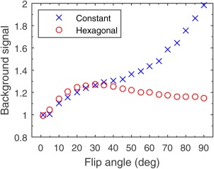
Figure 4.

RMS background intensity of in vivo liver spoiled‐gradient‐echo images for flip angles between 1 ° and 90 °, normalized to the same measure in a noise reference image. [Color figure can be viewed in the online issue, which is available at wileyonlinelibrary.com.]
