
Figure 1.
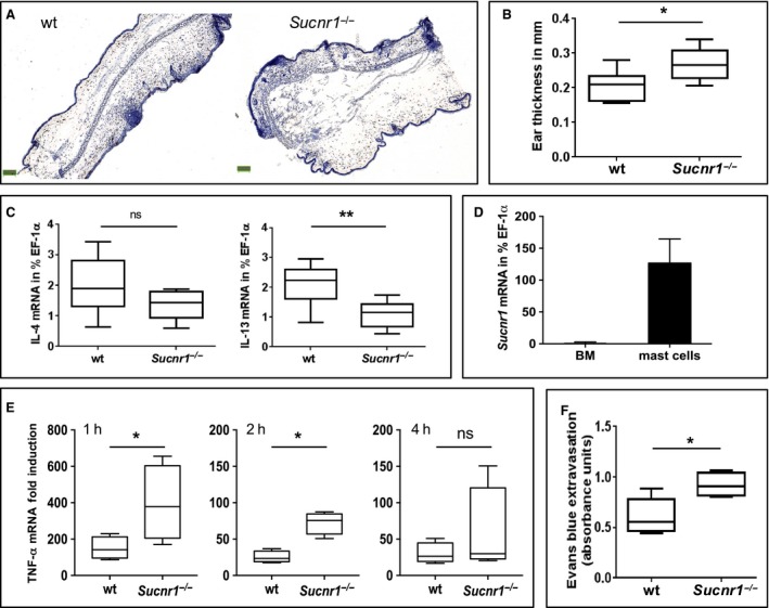
Sucnr1 −/− mice display exacerbated mast cell responses in vitro and in vivo. (A) Representative hematoxylin staining of ear sections from wt and Sucnr1 −/− mice after oxazolone challenge. (B) Comparative ear thickness from eight wt and eight Sucnr1 −/− oxazolone‐challenged mice. (C) Cytokine mRNA expression in oxazolone‐challenged ears from eight wt and eight Sucnr1 −/− mice. (D) Sucnr1 mRNA expression from bone marrow (BM) cells and BM‐derived mast cells isolated from two and eight, respectively, different mice. (E) TNF‐α mRNA expression by BM‐derived mast cells from wt and Sucnr1 −/− mice stimulated for indicated period of time with 2 μg/ml IgE plus 0.1 μg/ml DNP‐BSA. Boxplots represent fold increases in TNF‐α mRNA by stimulated mast cells related to corresponding unstimulated controls (pooled data from three independent experiments). (F) Evans Blue extravasation after passive cutaneous anaphylaxis (PCA) of five wt and five Sucnr1 −/− mice. Analysis using one‐tailed Mann–Whitney U‐test.