
Figure 5.
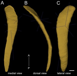
3D reconstruction of the mouse claustrum, shown in medial view (A), dorsal view (B), and lateral view (C). A‐P: Anterior to posterior direction. The dark blue lines depict anterior–posterior, medial–lateral, and dorsal–ventral axes in 3D. The mouse claustrum stretches for approximately 2.9 mm along the anterior–posterior axis, with a volume of 0.275 mm3. Scale bar = 250 μm.