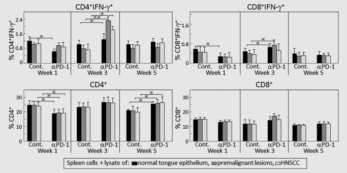
Figure 5.

PD‐1 antibody treatment transiently increases expression of IFN‐γ by CD4+ cells in response to lysates of premalignant oral lesion or HNSCC tongue tissues. Mice bearing 4NQO‐induced premalignant oral lesions were treated with either isotype control or PD‐1 antibodies. After 1, 3 and 5 weeks of treatment, sample mice were sacrificed and their spleens were cultured with lysates of control normal tongue tissue or lysates of premalignant lesion or HNSCC tongue tissues. Cultures were then immunostained for CD4, CD8 and IFN‐γ. Shown are percentages of positive‐staining cells from 5 mice per group per time point, with spleen cells from each mouse being cultured and phenotyped individually (mean ± SD). ✶=p < 0.05; ✶✶=p < 0.02.
