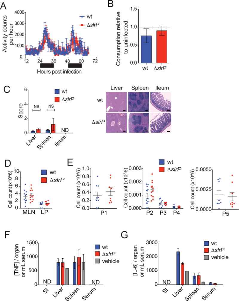
Figure 3. Canonical mediators of sickness-induced anorexia are equivalent between wt and ΔslrP ST.

(A) Activity of B6 mice orally infected with wt or ΔslrP ST. Black bar indicates dark/night cycle, and white bar indicates light/day cycle. n=6 mice/group.
(B) B6 mice were infected i.p. with wt or ΔslrP ST, and food consumption was measured every 24hrs post-infection and normalized to consumption of uninfected animals. Grams of food per animal consumed within the first four days of infection shown. n=6/group.
(C) H&E staining and histological scoring of spleen, liver, and ileum 24hr after oral infection of B6 mice with wt or ΔslrP ST. ND indicates pathology above limit of detection not detected. NS indicates not significant. Scale bar indicates 200μm. n=5/group.
(D) Total cellularity of MLN and SI LP 48hr after oral infection of B6 mice with wt or ΔslrP ST. n=12–13/group. Data represent 3 independent experiments combined.
(E) Numbers of inflammatory cell populations 48hr after oral infection of B6 mice with wt or ΔslrP ST. LP: T/B lymphocytes (P1, n=8–9/group), neutrophils (P2, n=13/group), phagocytic macrophages (P3, n=8–9/group), inflammatory monocytes (P4, n=8–9/group); MLN: migratory DCs (P5, n=8/group). Data represent 3 independent experiments combined.
(F–G) ELISA measurements of TNFα (F) and IL-6 (G) in indicated tissues 48hr after oral infection of B6 mice with wt or ΔslrP ST. n=4–10/group. ND denotes Not Detected. Vehicle indicates PBS. Error bars indicate +/− SEM. See Figure S2.
