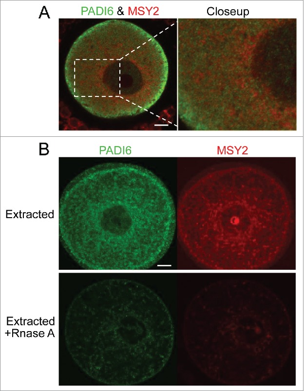
Figure 4.

PADI6 and MSY2 localize adjacent to each other in the normal oocytes and Triton extraction followed by RNase A treatment induces the loss of PADI6 and MSY2 from the oocytes. (A) Representative confocal images of PADI6 (green) and MSY2 (red) in the normal oocytes. (B) Representative confocal images of Padi6 WT oocytes stained with PADI6 (green) and MSY2 (red). All images parameters are the same in both conditions. Scale bar, 10 μm.
