
Figure 5.
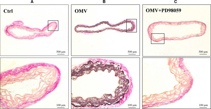
Immuohistochemistry for OMV‐induced VSMC calcification in mouse aorta. (A) After 3 weeks of aorta ring culture with PBS, aortic sections were stained using silver nitrate to detect calcium deposits. (B) Aorta rings cultured in the stimulation of 10 ng·mL−1 OMV for 3 weeks. (C) Aorta rings were cotreated with 10 ng·mL−1 OMV and ERK inhibitor. Representative images from three independent experiments are performed in duplicate. Higher magnification images of the boxed areas are shown under each image.