
Figure 4.
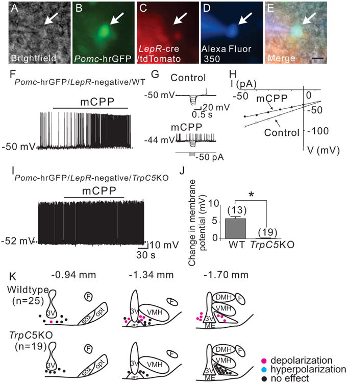
Trpc5 subunits are required for the acute mCPP-induced depolarization of arcuate Pomc neurons. (A) Brightfield illumination of Pomc-hrGFP neuron from PLT mice. (B) and (C) The same neuron under FITC (hrGFP) and Alexafluor 594 (tdtomato) illumination. (D) Complete dialysis of Alexa Fluor 350 from the intracellular pipette. (E) Merge image illustrates colocalization of hr-GFP and Alexa Fluor 350 indicative of a Pomc neuron which does not express Leprs. (F) Electrophysiological study demonstrates a Pomc-hrGFP (green) neuron from PLT mice that depolarized in response to mCPP (4μM). Traces showing decreased voltage deflection and increased action potential frequency after mCPP application. (H) Current versus voltage (I-V) plot from same WT neuron illustrating a characteristic decrease in input resistance subsequent to mCPP application. Shown are responses before (control) and during mCPP application. (I) demonstrates a current clamp recording of a Pomc-hrGFP∷Trcp5 -/Y (green) neuron in which mCPP fails to induce a depolarization. (J) Histogram summarizing the acute effect of mCPP on the membrane potential of Pomc neurons which do not express leptin receptors as well as express or do not express Trpc5 subunits (n= 13-19 per group). (K) Rostro-caudal and medio-lateral distribution of electrophysiological responses to mCPP from Pomc neurons which do not express leptin receptors as well as express or do not express Trpc5 subunits.