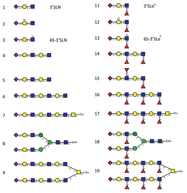
Figure 3.

Glycan structures of influenza A viruses. Structures are shown for sialylated glycans present in the array in nonfucosylated (glycans 1–9) and fucosylated (glycans 11–19) forms and binding by hemagglutinins is shown in Figures 2 and 7. Glycans 1 and 11 correspond to 3′SLN (nonfucosylated glycan) and 3′SLeX (fucosylated form of 3′SLN), respectively. Similarly, glycans 3 and 13 correspond to 6-O-sulfo 3′SLN (6S-3′SLN) and 6-O-sulfo 3′SLeX (6S-3′SLeX), respectively. Blue squares, N-acetylglucosamine; yellow circles, galactose; green circles, mannose; purple diamonds, sialic acid; red triangles, fucose. H5N12.3.4, novel H5N1 virus clade 2.3.4.
