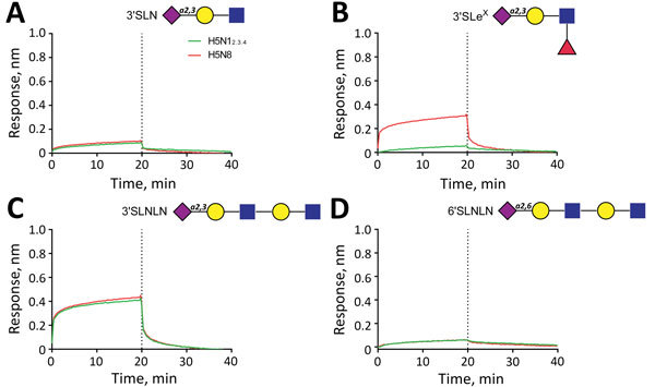
Figure 4.

Analysis of binding of influenza A virus H5N12.3.4 and H5N8 hemagglutinins to sialylated glycans by using biolayer interferometry. A) 3′SLN, B) 3′SLeX, C) 3′SLNLN, D) 6′SLNLN. After complexing biotinylated glycans with streptavidin sensors, association and subsequent dissociation of H5 proteins complexed with StrepMAB-classic (IBA GmbH, Göttingen, Germany) was determined. Blue squares, N-acetylglucosamine; yellow circles, galactose; purple diamonds, sialic acid; red triangles, fucose. The dotted lines at the 20-min time points distinguish the association and dissociation phases. H5N12.3.4, novel H5N1 virus clade 2.3.4.
