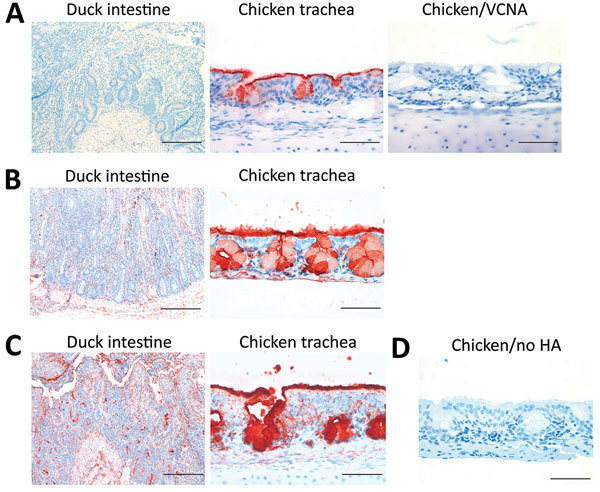
Figure 9.

Histochemical analysis of binding of influenza A virus H5 proteins to avian tissues. A) Duck intestine and chicken trachea tissues stained with an antibody specific for 3′SLeX (anti-3′SLeX). Tissue sections treated with Vibrio cholerae neuraminidase (VCNA) before immunostaining were used as controls. Scale bars indicate 200 μm in left panel and 50 μm in center and right panels. B, C) Duck intestine and chicken trachea tissues incubated with H5 proteins H5N12.3.4 and H5N8 after precomplexing with horseradish peroxidase (HRP)−conjugated antibodies. Scale bars indicate 200 μm in left panel and 50 μm in right panel. D) Chicken trachea tissues incubated with HRP-conjugated antibodies against H5N12.3.4 (no hemagglutinin [HA]) were used as a negative control. H5N12.3.4, novel H5N1 virus clade 2.3.4. Scale bar indicates 50 μm.
