
Figure 4.
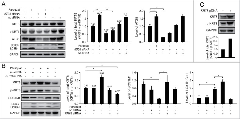
KRT8 expression is related to autophagy progression in RPE cells under oxidative stress. Western blot analysis of ARPE-19 cells after paraquat (400 μM) treatment of 24 h in the absence or presence of either (A) anti-ATG5 siRNA or (B) anti-KRT8 siRNA. Bar graphs indicate each protein expression level or the total KRT8 expression level (KRT8 + p-KRT8), and the numbers on the top of the quantitative bar graph represent the p-KRT8:total KRT8 ratio. Each protein band intensity was normalized to GAPDH. (C) Western blot analysis of KRT8 and its phosphorylated form (p-KRT8) in ARPE-19 cells overexpressing ARPE-19 and KRT8. Bar graphs show KRT8 and p-KRT8 expression levels normalized to GAPDH in western blot analysis.