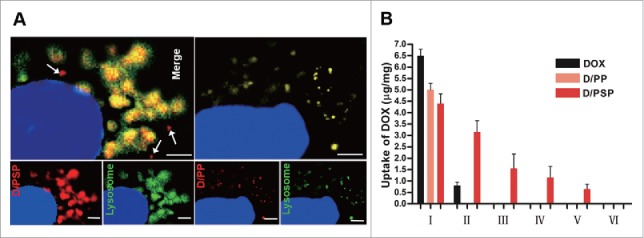
Figure 4.

Endo-lysosomal swelling effect and cellular uptake of PSP. (A) The appearance of D/PSP (left) and D/PP (right) in endo-lysosomes observed by CLSM. Endo-lysosomes were stained with LysoTracker Green, while the D/PSP was traced by red DOX. The white arrows indicate the PSP that escaped from endo-lysosomes. The nuclei were stained with Hoechst 33342. Scale bar: 2 μm. (B) Cellular uptake of DOX within cells during sequential intercellular delivery.
