
Figure 5.
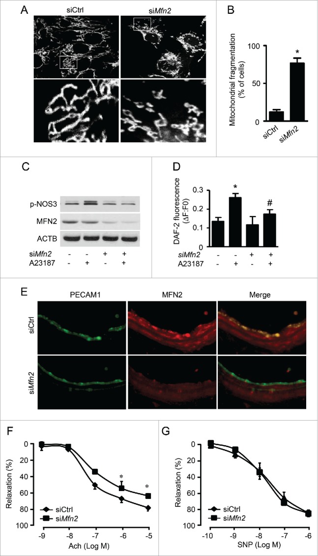
Inhibited mitochondrial fusion impairs endothelial function. (A) HUVECs were transfected with control (siCtrl) or MFN2 siRNA (siMFN2) for 48 h, and mitochondria were labeled with MitoTracker® Deep Red FM. Mitochondrial morphology was analyzed using fluorescence microscopy. Scale bars: 5 µm. (B) Mitochondrial fragmentation was determined, as described in Materials and Methods. n ≥ 100; *P < 0.05 vs. siCtrl. (C) HUVECs were transfected with siCtrl or siMFN2 for 48 h and then treated with A23187 or DMSO for 30 min. Phosphorylation of NOS-3 was measured by western blotting. (D) Nitric oxide concentration in the cells was assessed using the DAF-2 fluorescence probe. *P < 0.05 vs. DMSO; #P < 0.05 vs. siCtrl. (E) WT mouse carotid arteries were transfected with siCtrl or siMfn2 using electroporation for 48 h, stained with antibodies against MFN2 (Red) and PECAM1 (Green), and observed using fluorescence microscopy. (F) Endothelium-dependent vasodilator responses were measured in the presence of Ach (10−9 to 10−5 M). (G) Endothelium-independent vasodilator responses were measured in the presence of SNP (10−10 to 10−6 M). n = 4, * P < 0.05 vs. siCtrl.