
Figure 8.
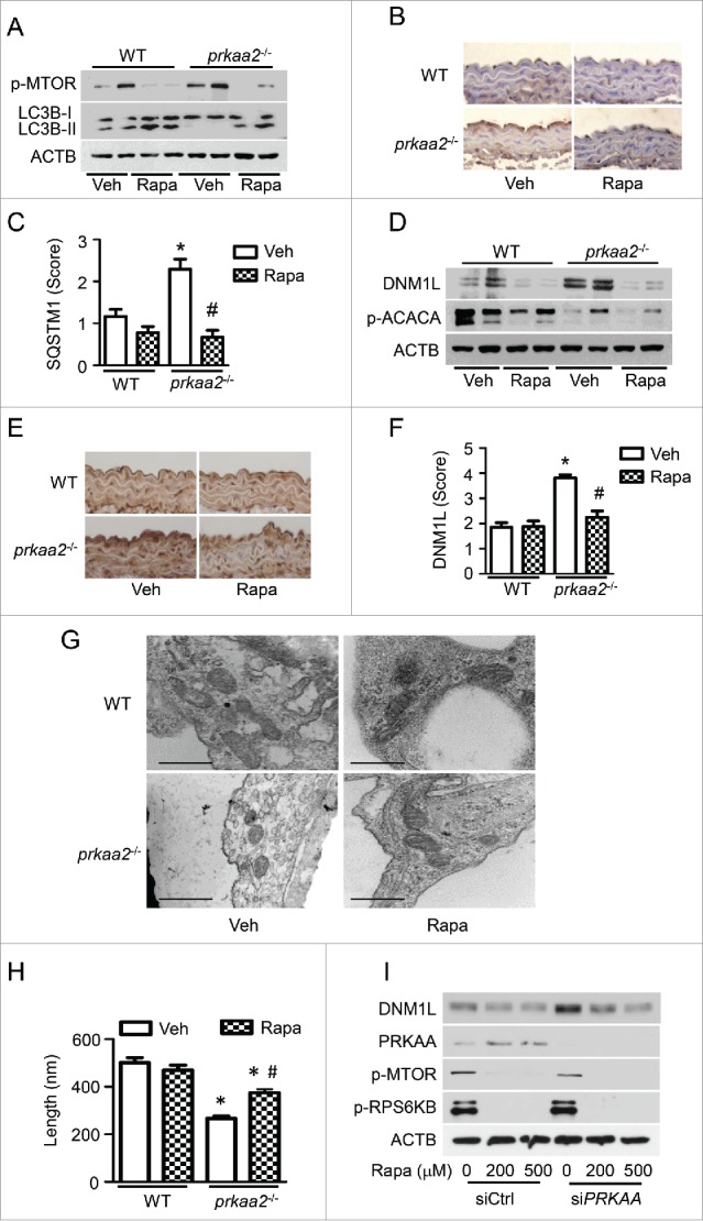
Activation of autophagy reduces DNM1L expression and mitochondrial fission in prkaa2−/− mice. WT and prkaa2−/− mice were orally administrated with rapamycin (Rapa, 14 mg/kg in diet) for 1 week. (A) Protein levels of phosphorylated MTOR at Ser2481 (p-MTOR) and LC3B in aortas were measured by western blotting. (B and C) Immunohistochemical staining for SQSTM1 in aortas from rapamycin-treated WT and prkaa2−/− mice, and quantification of positive staining for SQSTM1 in the aortas. *P < 0.05 vs. WT; #P < 0.05 vs. Vehicle. (D) Western blot analysis of DNM1L and p-ACACA protein expression in mouse aortas. (E and F) Immunohistochemical staining for DNM1L in aortas from rapamycin-treated WT and prkaa2−/− mice. Positive staining for DNM1L was quantified, as described in Materials and Methods. *P < 0.05 vs. WT; #P < 0.05 vs. Vehicle. (G) Representative electron micrographs of aortic endothelial mitochondria. Scale bars: 500 nm. (H) Mitochondrial length was quantified, as described in Materials and Methods. n = 6 mice, *P < 0.05 vs. WT, #P < 0.05 vs. Vehicle. (I) HUVECs were transfected with control (C-si) or siPRKAA for 24 h, after which they were treated with different concentrations of rapamycin for 24 h. Cell lysates were subjected to western blot analysis for DNM1L, PRKAA, p-MTOR, and phosphorylated RPS6KB at Thr389 (p-RPS6KB).